• Hyperion X5 3D PAN Ceph

      MyRay - Italy
      1 2 3 4 5
      Availability:
      С предварителна поръчка

      3D Multi FOV imaging system with Focus-Free PAN designed for all users and factory-set for upgrading at any time with a teleradiographic application. Designed to simplify dental diagnostics with 3D and 2D images that can be viewed in real time.

      Hyperion X5 3D PAN CEPH

      3D Multi FOV imaging system with Focus-Free PAN and Full CEPH accessible for all users, suitable for wall mounting. Designed to make complete dental diagnostics accessible in real time.

      None

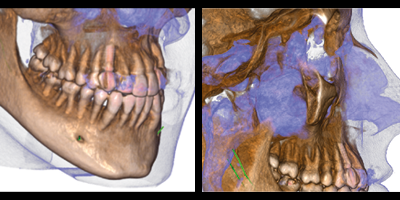
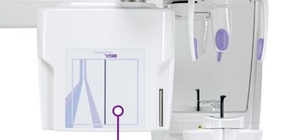
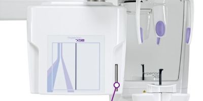
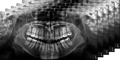
      Focus-free 2D with MRT.

      The PAN examination uses MRT (Morphology Recognition Technology) and an automatic best focusing selection system. A multi-layer panoramic scan is performed, with automatically optimised exposure and scan times for children and adults.

      None

      Cephalometric examination.

      The renewed Hyperion X5 Ceph teleradiographic system provides programmes for every diagnostic need. Ultra-high quality images, extremely short scan times and low radiated doses: the very best cephalometric technology, all in the most compact unit the market has to offer.

      None

      High resolution CB3D.

      HD 3D imaging with ultra-fast, low-dose scans and very high resolution: 80 μm over the complete dentition,together with dedicated FOVs developed to ensure the best imaging at all times. Complete dental diagnosis, including assessment of maxillary sinuses.

      All the potential of 3D.

      Accessing the potential of 3D exams has never been easier or more effective. Thanks to dedicated mechanisms, patient positioning solutions and exclusive automatisms that help ensure a positive outcome with every examination, dentists can exploit the full potential of 3D.

      • Automatic sensor and collimator alignment
      • Ultra-high sensitivity 3D-PAN sensor
      • Adjustable and ergonomic head support
      • 3D MultiFOV, from 6x6 to 10x10 cm
       
      None

      3D - PAN SENSOR

      The high-sensitivity 3D sensor is also versatile as it can perform 2D panoramic exams (managed by programmes in the software package and controlled via the user-friendly virtual control panel).

      None

      AUTOMATIC CEPH COLLIMATION

      With cephalometric examinations the turret containing the 3D sensor automatically rotates and descends, aligning so its opening-equipped structure creates the collimation suitable for the examination. Moreover, the sensor is positioned so there is more space for the patient and the experience is a more comfortable one.

      None

      HEAD SUPPORT WITH 5 CONTACT POINTS

      The dedicated head support for volumetric examinations has 5 contact points. Three of these - frontal, right and left - are adjustable. This improves patient positioning and, consequently, stability and clinical examination quality.

      A wide range of FOVs

      A wide range of FOVs available for your clinical needs: from implantology to the measurement of maxillary sinus volumes, from endodontics to oral surgery. Each FOV is available in three versions to adapt to all clinical needs. It takes just a few simple steps to identify the most suitable set-up based on the anatomical region of interest.

      • QuickScan Faster and ultra-low dose scans for post-surgery follow-up and macrostructure analysis.
      • Standard mode Primary diagnostics and treatment planning. The best balance between dosage and quality.
      • SuperHD Outstanding, uncompromising level of detail. Ideal for micro-structure analysis.


      FOV 10X10

      FOV 10X10

      FOV 10X6 e 10X7

      FOV 10X6 e 10X7

       
      Broaden your vision, expand your diagnosis: in a single scan, Hyperion X5 can show you the entire dentition, including third molar roots or maxillary sinuses of adult patients, via ultra-fast (6.4 s) scans at ultra-low doses, or with very high resolution up to 80 μm  

      Fields designed for lower arch imaging including third molars, and upper arch imaging including the maxillary sinus floor. Maximum amount of information in a single volume, for more complete case studies.


      FOV 8X6 e 8X7

      FOV 8X6 e 8X7

      FOV 6X6 e 6x7

      FOV 6X6 e 6x7

      Reduced fields of view suitable for examining the upper or lower semi-arch in adult patients, or with limited doses for the examination of the complete dentition in children. 6 cm diameter to view sections along the dental arch. It only scans your area of interest: semi-arches or frontal areas, without cutting out the occlusal zone or the lower base of the upper jaw and minimising patient exposure.


      CONTROL VIA iPAD

      Hyperion X5 has a user-friendly graphical interface, also available in the iPad application. It promotes intuitive control: in a few simple steps you can choose and set up the most appropriate examination based on clinical and anatomical interest.

      None

      PC INTERFACE

      The multi-platform console allows simple and immediate access to all the device’s features. The interface guides you step by step through every stage, from examination selection to set-up, with guided positioning of the FOV: for easier, faster and more effective scanning


      CARING FOR WELL-BEING.

      Hyperion X5 simplifies your work and promotes the well-being of your patients. Quick scans, ultra-low dose irradiation, procedures that contribute to creating a peaceful and collaborative environment. Easy for you, comfortable for your patient.

      None

      EFFECTIVE GUIDED POSITIONING

      Positioning is fast and accurate thanks to an alignment system that projects 3 laser beams directly on to the patient’s face, and the ergonomic head support unit equipped with 4/5 contact points ensuring the highest stability during scanning. The large mirror helps positioning while allowing maximum freedom of movement. The patient will always feel at ease.

      None

      SERVO-CONTROLLED SYSTEM

      The Scout View system allows the volume to be centred on the area of interest, keeping the patient in the same comfortable position. From the PC, the operator can see two (sagittal and frontal) views at ultra-low dose irradiation and fine-tune the scanning area, allowing the equipment to reposition itself correctly with very precise servo-assisted movements. This procedure avoids having to repeat the examination.


      ULTRA LOW DOSE QUICK SCANNING

      The advanced QuickScan protocols, available for both 2D and 3D examinations, allow accurate images to be obtained at lower doses compared to standard image acquisition. They are the ideal tool for post-operative monitoring and the identification of any macro-structures (such as impacted teeth and agenesis).


      iRYS, simple and versatile diagnoses.

      The all-in-one software designed for simple and effective management of 2D and 3D images, with advanced tools and filters for diagnostics and planning.

      Equipped with a whole ecosystem of features to view and process data captured during examinations, iRYS makes the diagnostic process easier and helps share images directly from a dedicated workstation to the dental surgery computers and the iRYS Viewer application available for iPAD. With just one click you can send 2D images and 3D volumes to dental practice management software or to advanced design systems (guided implantology, cephalometric tracking, etc.).

      • Multi-desktop 2D/3D
      • Simplified implant libraries
      • Bone quality assessment
      • Airway volume analysis
      • IRYS Viewer dynamic reporting (APP for iPAD)